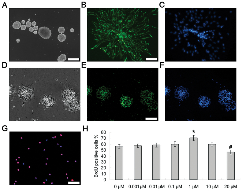
Figure 1.
Fluoxetine increased the proliferation of NPCs. (A) Typical neurosphere morphology of rat embryonic neural precursor cells maintained in growth medium. (B–C) Immunostaining of Nestin (green) and DAPI (blue) in NPCs. Scale bars = 20 μm. (D–F) Immunostaining of sox2 (green) and DAPI (blue) in NPCs. (G) For cell proliferation, NPCs were incubated for 2 d in the presence of increasing concentrations (0–20 μM) of fluoxetin. Values represent means ± standard deviation (n = 5). BrdU-positive cells and nuclei (DAPI) were labeled with red and blue. Scale bars = 20 μm. (H) Quantification of data. ANOVA revealed a main effect of treatment [F (5, 24) = 9.67, p < 0.0005]. *p < 0.01 versus control (0 μM); #p < 0.005 versus control. BrdU, 2 d, 5’-bromo-2-deoxy-uridine; DAPI, 4,6-diamidino-2-phenylindole; NPCs, neural precursor cells.

