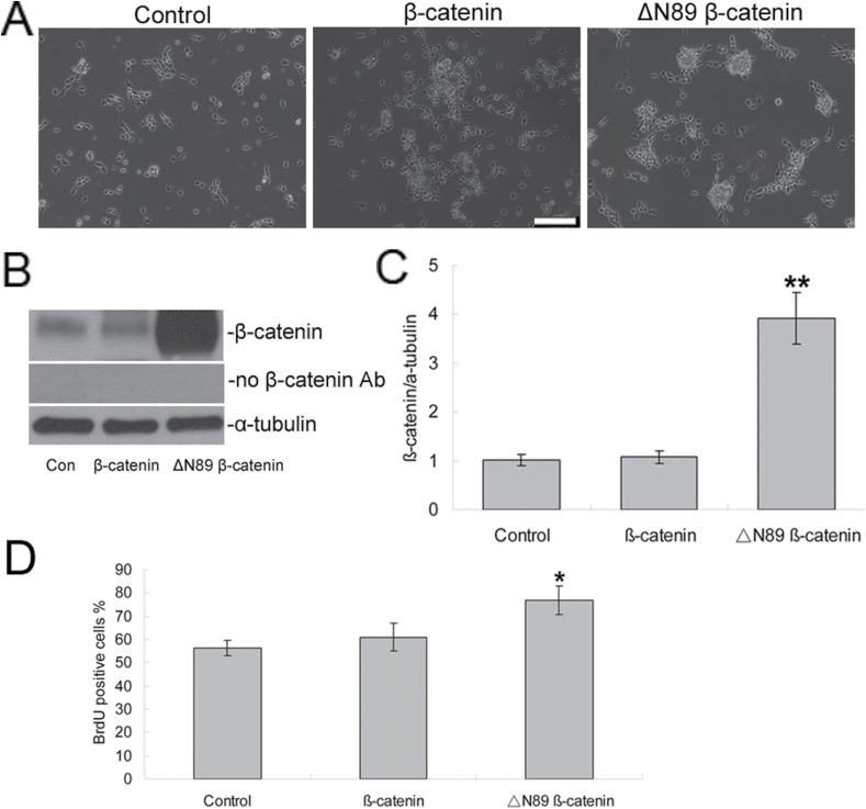
Figure 5.
Transduction of stabilized β-catenin increased the proliferation of NPCs. NPCs were transduced with pSin-EF2-beta-catenin, pSin-EF2-deltaN89 beta-catenin, and pSin-EF2-GFP. (A) Phase contrast images of NPCs after transduction for 48h. Scale bars = 20 μm. (B) The protein expression of β-catenin was further detected by Western blotting. (C) Quantification of Western blotting signals of β-catenin and α-tubulin proteins. Data were ratios compared with α-tubulin protein. n = 5 for each group. **p < 0.005 compared with the control group. (D) 48h after transduction, cell proliferation was measured by BrdU labeling. Values represent means ± standard deviation (n = 5). *p < 0.01 versus the control group. BrdU, 2 d, 5’-bromo-2-deoxy-uridine; NPCs, neural precursor cells.

