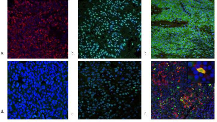
Fig. 1.
Demonstration of dual corticotroph (assessed with ACTH, Tpit, and NeuroD1 markers) and gonadotroph (assessed with LH, DAX-1, and SF-1 markers) lineage in an SCA. Fluorescence immunohistochemistry of corticotroph markers ACTH, Tpit, NeuroD1, LH, DAX-1, and SF-1 were performed in a silent corticotroph adenoma. Tumor was fixed in 10% formalin, paraffin embedded, and stained with primary antibodies, with Alexa 488 secondary antibodies and visualized with confocal immunofluorescence microscopy at 20-40 x magnification. Tumor is (a) ACTH positive (b) NeuroD1 positive (c) SF-1 positive (d) Tpit negative, and (e) DAX-1 positive. (f) Tumor exhibits co-localization of ACTH and LH, showing potential derivation of SCAs from dual corticotroph-gonadotroph line. Portion of the field enlarged for detail (100 x) highlights the co-localization in a cell. [10]

