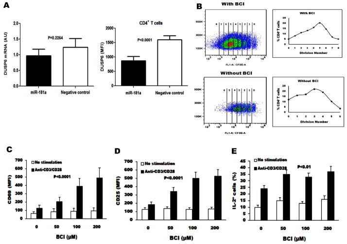
Fig. 5. miR-181a regulates DUSP6 gene expression at the translational level and DUSP6 inhibition augments T cell activation.
A) The levels of DUSP6 mRNA in CD4+ T cells from HCV-infected individuals with transfection of miR-181a precursor and negative control were analyzed by real-time PCR (left panel), and DUSP6 protein expression by flow cytometry. DUSP6 expression was shown as mean fluorescent intensity (MFI) in CD4+ T cells transfected with miR-181a precursor compared with those transfected with negative control (right panel). B) Purified CD4+ T cells from 6 HCV-infected individuals were labeled with CFSE, stimulated with anti-CD3/CD28 in the presence of IL-2 and (E)-2-benzylidene-3-(cyclohexylamino)-2, 3-dihydro-1H-inden-1-one (BCI) ex vivo for 5 d, and followed by flow cytometry analysis. Representative dot plots of cell division as CFSE-dilution in the gated CD4+ T cells from BCI treated and non-treated are shown here. C–E) Purified CD4+ T cells from HCV-infected individuals with anti-CD3/CD28 antibodies in the presence of increasing concentrations of BCI. The MFI of CD69, CD25, and IL-2 expressions were analyzed by flow cytometry and shown as mean±SE from 6 HCV-infected individuals.

