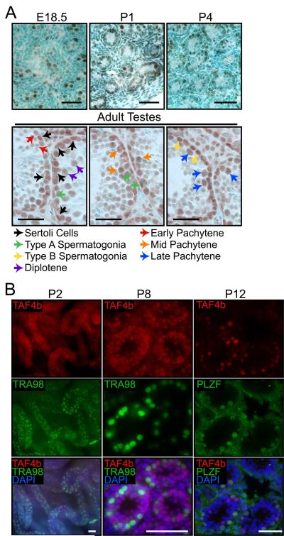
Figure 1. Germ and somatic cell TAF4B protein enrichment during testes development.
(A) Immunohistochemistry analysis of TAF4b protein localization during late embryonic, early postnatal and adult testes sections. Corresponding colored arrows indicate examples of TAF4b protein enrichment in different somatic and germ cell types. Scale bars = 50 μm. (B) P2 testes whole mount and P8 testes section immunofluorescence show colocalization of TAF4b (red) with TRA98 (green) in gonocytes, as well as TAF4b enrichment in TRA98-negative somatic cells. P12 testes sections show TAF4b protein enrichment (red) in both PLZF-positive (green) and PLZF-negative spermatogonia. DAPI (blue). Scale bars = 50 μm.

