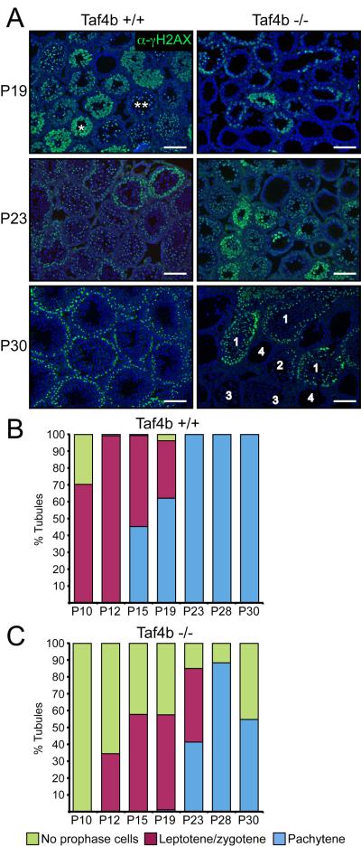
Figure 4. Delayed meiosis initiation and progression in Taf4b −/− testes.
(A) γH2AX immunofluorescence (green) of P19, P23 and P30 testis sections from wild type and Taf4b −/− mice. P30 Taf4b −/− seminiferous tubule phenotypic categories: (1) normal, (2) partial absence of spermatocytes, (3) having spermatids undergoing spermiogenesis but lacking spermatocytes, (4) completely devoid of meiotic spermatocytes and spermatids. DAPI (blue) * Denotes leptotene/zygotene stage spermatocytes with diffuse γH2AX staining, ** denotes pachytene stage spermatocytes with XY-body γH2AX foci. Scale bars = 100 μm. Quantitative analysis of γH2AX staining and meiotic progression during the first wave of spermatogenesis in wild type (B) and Taf4b −/− (C) testes. Tubules containing multiple meiotic cell types are categorized by furthest stage. Between 100 and 200 tubules were counted for each data point.

