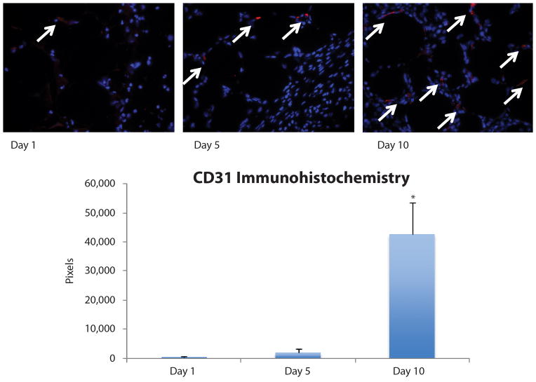
Figure 5.
CD31 Immunohistochemical staining. Increased graft vascularization was detected by CD31 immunohistochemical staining (red pixels highlighted by white arrows) at day 10 (426,148 ± 189,071 pixels) compared to day 1 (3,249 ± 5,628 pixels, *p < 0.05) and day 5 (18,104 ± 26,679 pixels, *p < 0.05) post-grafting. Vascularity at day 5 was higher than day 1, but the difference did not reach statistical significance (p > 0.05).

