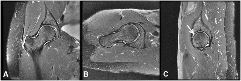
Figure 2.

Examples of clinical images acquired on the right hip of a 52 year old female subject with KL=2 and the presence of acetabular and cartilage lesions. The images represent (a) the coronal FSE (b) the axial FSE, and (c) sagittal FSE acquisitions. An arrow on the sagittal FSE points to the location of an acetabular cartilage lesion.
