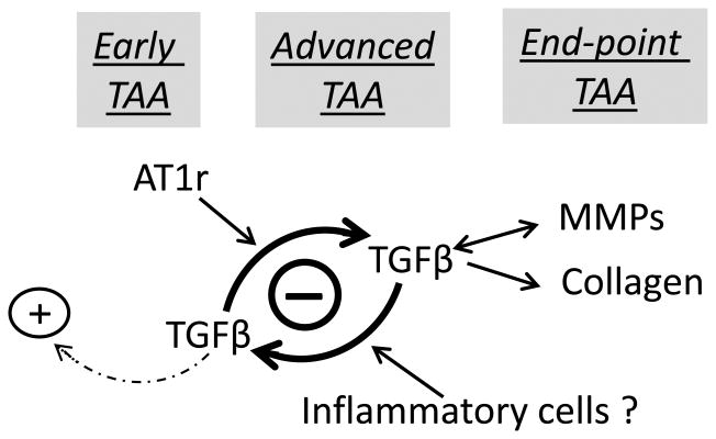
Figure 6. Proposed new model of TAA pathogenesis in MFS.
Schematic representation of the progressive stratification of signaling events driving arterial disease during the early, advanced and end-point stages of TAA in mice with severe MFS. The model focuses on AT1R and TGFβ signaling and assumes that recurring hemodynamic load is the primary trigger of aorta dysfunction. The (+) and (-) symbols respectively highlight the protective and detrimental roles of baseline and overactive TGFβ signaling; the dotted arrow signifies the unproductive response of baseline TGFβ signaling to wall stress; and the double-headed arrow indicate the reciprocal pathogenic relationship between TGFβ and MMP hyperactivity.

