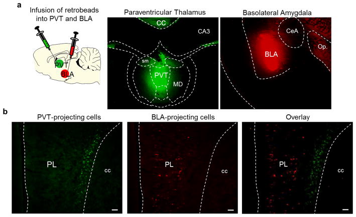
Extended Data Fig. 7. PL neurons projecting to PVT vs. BLA are located in distinct layers.
(a, left) Schematic of retrobead injections. (a, middle) Micrograph showing the site of retrobeads infusion into PVT (green) and BLA (red) (a, right) in the same rat. (b, left) PL neurons retrogradely labeled from PVT infusion (green). (b, middle) PL neurons retrogradely labeled from BLA infusion (red). (b, right) Overlay image showing absence of co-labeling between PL neurons projecting to PVT (green, deep layers) and PL neurons projecting to BLA (red, superficial layers). Scale bar, 100 μm.

