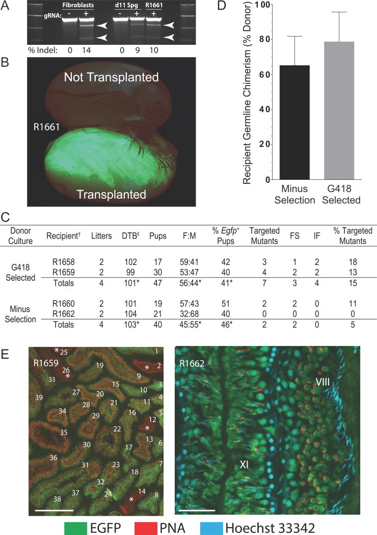
Figure 1. CRISPR/Cas9-Mediated Gene Targeting in Donor Stem Spermatogonia.
(A) CRISPR/Cas9 cleavage of Epsti1 in rat Fibroblast and Spermatogonial (Sg d11) cultures on d11 post-transfection, and in flow sorted EGFP+ spermatogenic cells (R1661) on d56 after transplantation into the right testis of recipient R1661: transfection without (−) and with (+) Epsti1 gRNA. Arrows: predicted ~461 bp + ~226 bp Surveyor products.
(B) Spermatogenesis (green fluorescence) in testis of R1661 on d56 after transplantation that developed from donor EGFP+ rat spermatogonial cultures containing CRISPR/Cas9- catalyzed Epsti1 mutations.
(C) Targeted mutagenesis of Epsti1 exon 2 in rats by CRISPR/Cas9 spermatogonial gene editing. Donor spermatogonia were hemizygous for tgGCS-EGFP. FS, frame shift mutation; IF, mutation in frame, DTB, days from transplantation to first litter containing Epsti1 mutant animals. *Average value, †R(n), Recipient identifier, ||Litters born 99 to 128 d post-transplantation, % of total F1 progeny/recipient. See also Figure S1.
(D) Donor germline chimerism (%) in wildtype recipient rat testes. Ratio of seminiferous tubules containing EGFP+ elongating spermatids/total tubules containing elongating spermatids plotted/donor culture condition (Minus Selection and G418 Selected). See also Table S1 and Table S2.
(E) Acrosome (PNA) & nuclear (Hoechst 33342) labeling marking donor-derived elongating spermatids (EGFP+) in tubule cross sections from recipient R1659. Left, 39 of 39 tubules contained elongating spermatids (EGFP+ or EGFP−). Five EGFP− tubules are marked with an asterisk. Scale 500 μm. Right, higher magnification image of donor spermatogenesis at stages VIII and XI in the rat seminiferous epithelial cycle [See: (Hess, 1990)]. Scale 100 μm. See also Figure S2.

