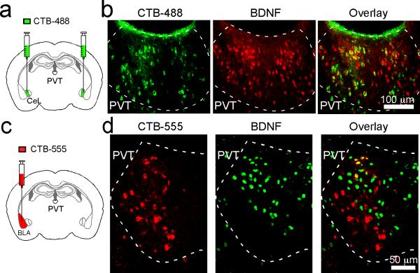
Extended Data Figure 6. CeL-projecting neurons in pPVT express BDNF.
a. A schematic of the experimental approach to retrogradely label CeL-projecting pPVT cells. b. Representative images of pPVT cells, which were labeled by CTB (left) and an antibody recognizing BDNF (middle). CTB-labeled neurons largely overlapped with BDNF-positive somatas (see overlay in right). (c, d) BLA-projecting neurons and BDNF-expressing neurons in pPVT are largely non-overlapping. c. A schematic of the method used to label BLA-projecting neurons in pPVT. d. Representative images of pPVT cells labeled by either CTB-555 (left) or the antibody recognizing BDNF (middle). These two populations were largely non-overlapping (see overlay in right).

