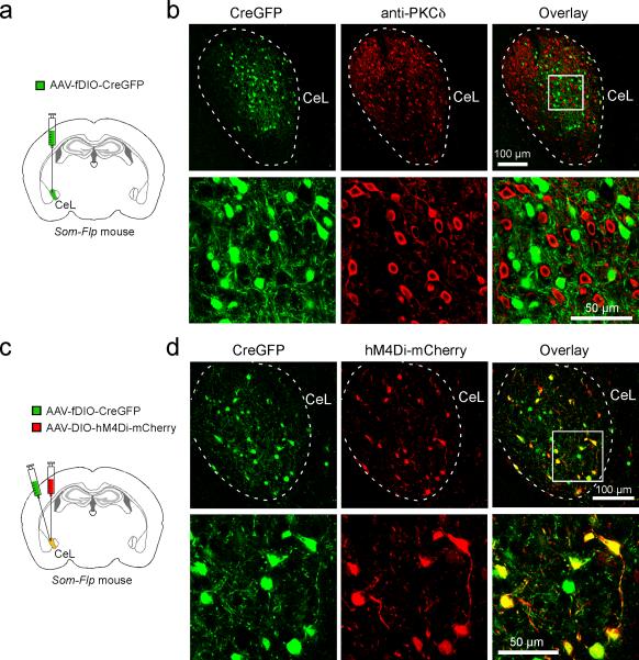
Extended Data Figure 8. Characterization of the AAV-fDIO-CreGFP.
a. A schematic of the experimental approach to selectively target SOM+ CeL neurons in the Som-Flp mice with the AAV-fDIO-CreGFP. b. Representative images of CeL neurons expressing CreGFP (left), and PKC-δ+ CeL neurons (as surrogate for SOM– neurons) that were recognized by an antibody (middle). In lower panel are high magnification images of the boxed region in the upper panel. These two cell populations were largely non-overlapping (see overlay on right), indicating that the AAV-fDIO-CreGFP selectively infects SOM+ neurons (data from one mouse). c. A schematic of the experimental approach to test the function of AAV-fDIO-CreGFP, whereby the CeL of Som-Flp mice was injected with a mixture of AAV-fDIO-CreGFP and AAV-DIO-hM4Di-mCherry. As the latter virus expresses mCherry in a Cre-dependent manner, observation of selective mCherry expression in GFP+ neurons would indicate that the AAV-fDIO-CreGFP is effective. d. Sample images of CeL neurons expressing CreGFP (left) and mCherry (middle). In lower panel are high magnification images of the boxed region in the upper panel. Essentially, all mCherry+ neurons co-expressed GFP (see overlay on right), indicating selective expression of Cre by the GFP-labeled cells (data from one mouse).

