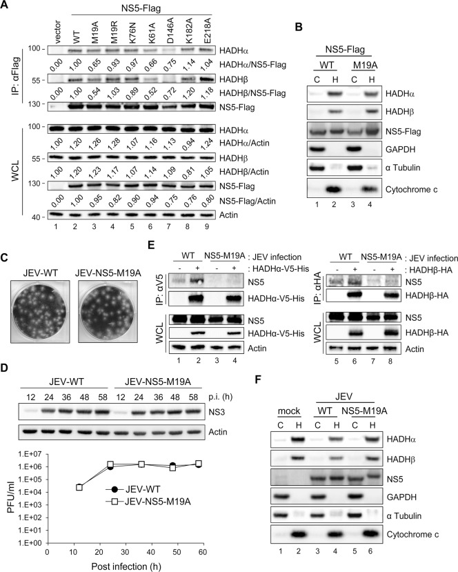
Fig 6. NS5 with mutation on residue 19 (M19A) showed reduced binding ability with MTP.
(A) Western blot analysis of indicated proteins in HEK293T cells transfected with the plasmids expressing wild type (WT)-, mutated-NS5-Flag or vector control for 24 h after immunoprecipitation with anti-Flag affinity gel. Band densities were quantified by use of MetaMorph (Molecular Devices). (B and F) HEK293T cells transfected with NS5-Flag or NS5-M19A-Flag (B) or infected with wild-type JEV (JEV-WT) or JEV-NS5-M19A (MOI = 5) (F) for 24 h underwent Qproteome Mitochondria Isolation. Western blot analysis of indicated proteins in mitochondrial and cytosolic fractions. C, cytosolic fraction; H, heavy membrane fraction/crude mitochondrial fraction. (C and D) JEV-NS5-M19A mutant virus was generated by using a JEV infectious clone. (C) Plaque morphology of wild type JEV (JEV-WT) and JEV-NS5-M19A mutant in BHK-21 cells. (D) A549 cells were infected with JEV-WT or JEV-NS5-M19A (MOI = 0.1) for the indicated times. Western blot analysis of protein levels of NS3 and actin. Plaque-forming assay of virus titration in culture supernatants (n = 3). Data are mean±SD. (E) IP analysis with V5 or HA affinity gel and Western blot analysis with the indicated antibodies in HEK293T cells adsorbed with JEV for 1 h, then transfected with HADHα-V5-His or HADHβ-HA for 24 h.

