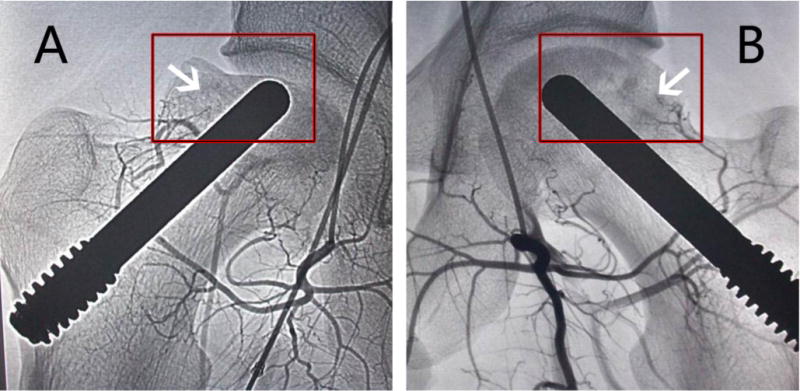
Fig. 4.

The photographs were taken by digital substraction angiography at 36 months. (A) Blood vessel regeneration was not found in the hip of the control group (arrow), and the femoral head collapsed (in box); (B) Blood vessel regeneration was observed in the hip of the combination treatment group (arrow), new blood vessels were developing sufficiently to reach the femoral head region (arrow), and the femoral head remained intact and round (in box).
