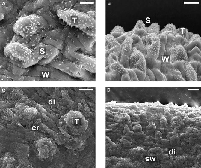
Fig 2. Scanning electron microscopy of the mid-body dorsal region of male S. mansoni worms recovered from a mouse 45 days post-infection by portal perfusion.
A and B: Untreated (control) worms. C and D: worms from rodents treated with 40 mg/kg EPI. Scales bars in A and C show 5 μm, and in B and D show 10 μm. (A): dorsal tegumental surface showing tubercles (T), apical spines (s) and parallel wrinkles (w). (B) Lower magnification of (A) showing the same features regularly appearing across surface. (C) and (D): SEM of male worm after treatment with EPI: Tubercles (T) and the lateral tegument disintegrate (di) resulting in disappearance of the knobs, spines and many of the parallel-arranged wrinkles. Dorsal tegumental surface shows swelling (sw), and erosion (er) of the surface with the exposure of subtegumental tissue. Overall appearance quite different to that of the control worms, due to erosion of tubercules and loss of all apical spines.

