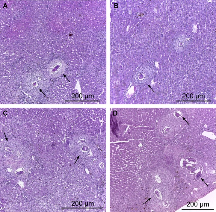
Fig 3. Photomicrographs of hepatic granulomas of mice liver 45 days post infection.
A and B: Liver of treated infected animals (40 mg/kg). Note the necrotic-exudative phase with loose inflammatory infiltrate (macrophages, neutrophils and eosinophils) and areas with necrotic cells around the egg. C and D: Liver of untreated infected animals, the granulomas were found in the periportal area. Different stages of evolution of the granuloma, including granulomatous pre-exudative, necrotic-exudative, productive and healing by fibrosis were observed. Arrows indicate the presence of inflammatory cells.

