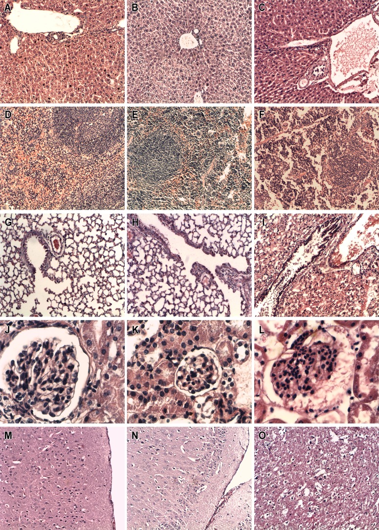
Fig 4. Histopathological study of organs sections of different groups of mice.
Comparison between the histology of: liver (A,B,C), spleen (D,E,F), lung (G,H,I), kidney (J,K,L), and brain (M,N,O) obtained from Swiss mice (n = 4) treated with 0.0 (first column), 530 (second column) or 8000 (third column) mg/kg of EPI. The first and the second column refer to mice killed 7 days after assay and the third one represent organs from animals that died due to the administration of higher concentration of the drug. Light microscopy evaluation of these organs showed severe morphological changes in the spleen, lung, kidney and brain, but not in the parenchyma of liver in the animals treated with 8000 mg/kg of EPI. Focal alterations in the red pulp were observed in the spleen of animals treated with 530 mg/kg of the drug. Control group presented no morphological changes in the organs.

