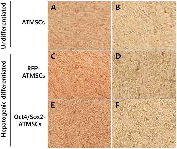
Fig 3. Morphology of RFP- and Oct4/Sox2-ATMSCs after 28 days hepatogenic differentiation.
(A,B) Undifferentiated ATMSCs showed fibroblast-like morphology without morphological changes. (C,D) Hepatogenically differentiated RFP-ATMSCs and (E,F) hepatogenically differentiated Oct4/Sox2-ATMSCs exhibited significantly changed morphology and developed a round or polygonal epithelioid shape during step-2 of differentiation. Statistical analysis was performed by student t-test (significant, **p < 0.01).

