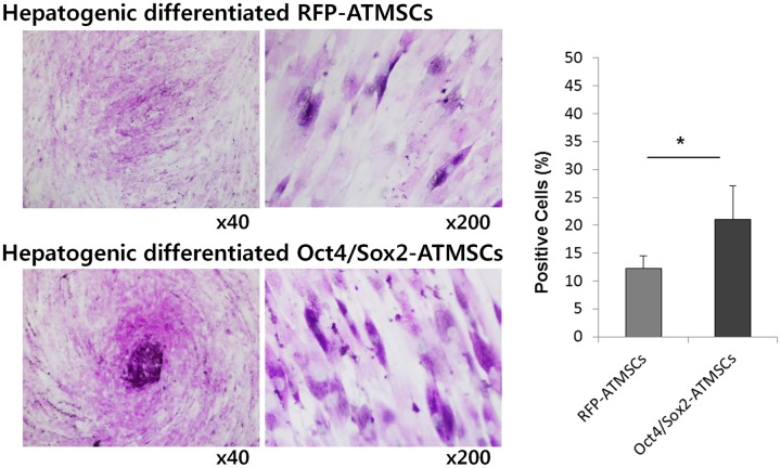
Fig 5. Period acid Schiff (PAS) staining of RFP- and Oct4/Sox2-ATMSCs after 28 days hepatogenic differentiation.
(A) Detection of glycogen in the cytoplasm of MSCs subjected to the liver differentiation protocol was demonstrated by PAS staining. PAS-positive substances stain pink in the cytoplasm of cells. (B) The number of PAS-positive cells is expressed as percentage of the total number of counted cells and was significantly higher in Oct4/Sox2-ATMSCs than that of RFP-ATMSCs. The experiments were repeated at least three times and similar findings were observed. Data represent the mean ± SD of three independent experiments. Statistical analysis was performed by student t-test (significant, *p < 0.05).

