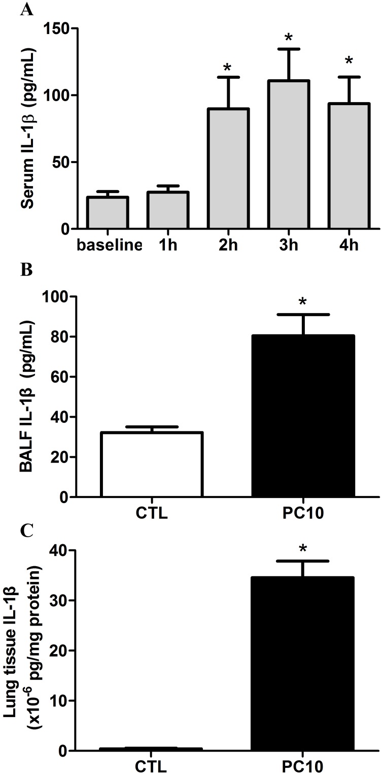
Fig 3. The IL-1β cytokine response in animals exposed to VILI.
A: in serum, B: in BALF, C: in lung tissue homogenization. The cytokine profiles of IL-1β from collected serum, BALF, and lung tissue homogenization samples, respectively, were shown by ELISA kit assay. A: Time course of IL-1β in serum, in animals exposed to 10 cmH2O pressure of ventilation. B and C: The expressions of IL-1β in BALF and lung tissue homogenization collected in animals exposed to 10 cmH2O of ventilation pressure for 4 hours. PC10: animals exposed to VILI via high pressure ventilation at 10 cmH2O for 4 hours, CTL: control group animals without ventilator administration. N = 8 per group, *: P <0.05 compared with baseline point or to animals without ventilator. The results are presented in terms of mean±SEM.

