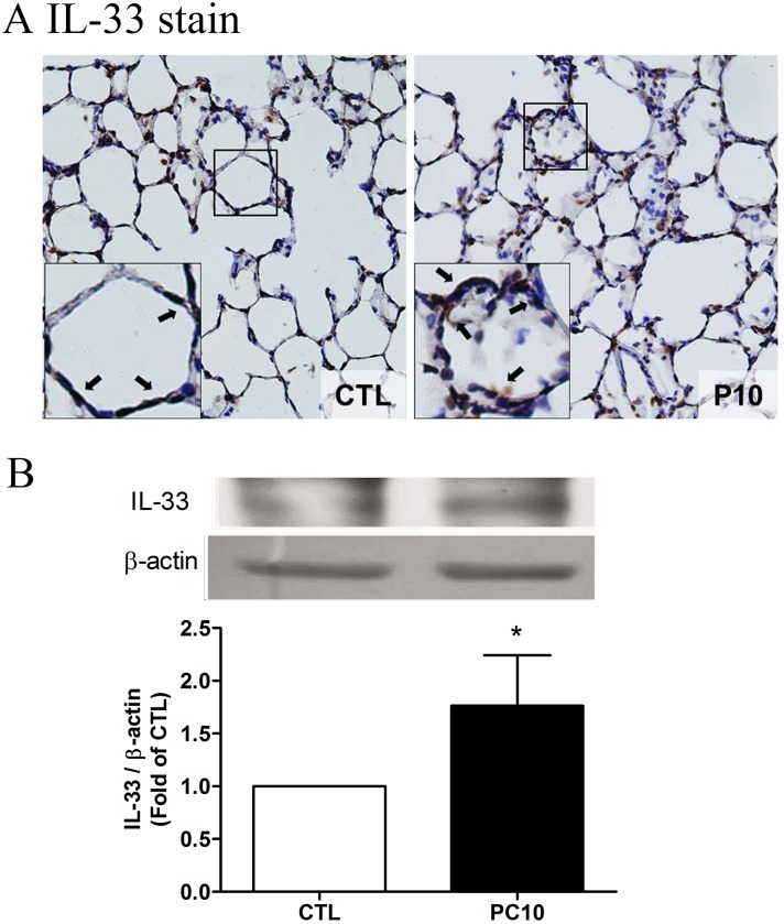
Fig 5. The IL-33 expressed in lung tissue in animals exposed to high pressure ventilation for 4 hours.
A: The IL-33 expression of lung tissue sections was determined using immunohistochemistry analysis and colorimetric detection with DAB (brown stain). The figure is presented at a magnification of 400X (insert, 1000X). The arrows reveal that IL-33 staining was remarkably accumulated in the alveolar wall. The slides were incubated using rabbit anti-IL-33 polyclonal antibody. B: IL-33 expression of lung tissue homogenization was determined using western blot assay and densitometry analysis. PC10: animals exposed to ventilation at 10 cmH2O for 4 hours, CTL: control group animals without ventilator administration.*:P <0.05 compared with animals without ventilator, n = 8. The results are presented in terms of mean±SEM.

