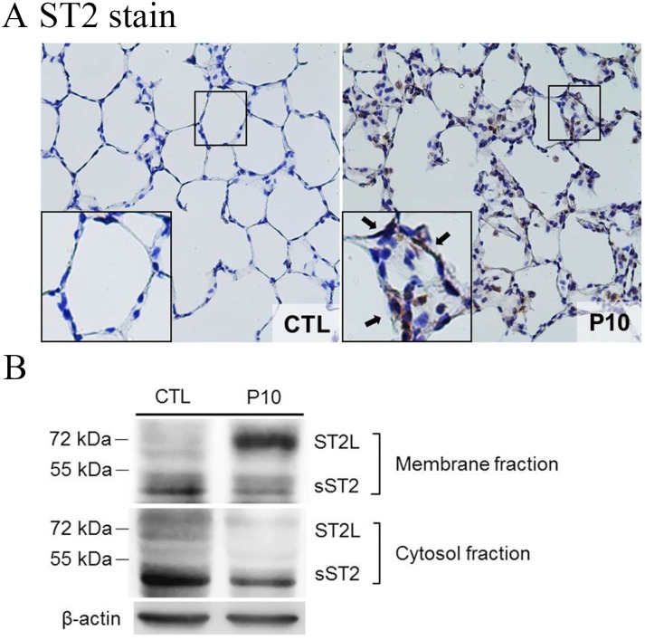
Fig 6. ST2 receptor translocated to cell membranes in lung tissue of rat during VILI.
A: The ST2 expression in lung tissue sections was analyzed by using IHC staining with rabbit anti-ST2 polyclonal antibody conjugated DAB (brown stain). The figure is shown at a magnification of 400X (insert, higher magnification of 1000X). The arrows indicate high accumulation of ST2 immunostaining in the alveolar wall. PC10: animals exposed to ventilation at 10 cmH2O for 4 hours, CTL: control group animals without ventilator administration. B: Lung tissue homogenization samples were separated into membrane and cytosol fractions and then subjected to western blot assay.

