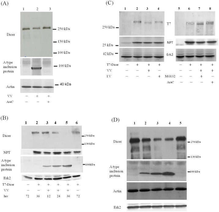
Fig 1. Reduction of Dicer protein during vaccinia virus infection.
(A) HeLa cells were either mock infected (lane 1) or infected with VV (M.O.I. = 10) in the absence (lane 2) or in the presence (lane 3) of AraC. Sixteen hrs after infection, cell lysates were analyzed by SDS-PAGE and Western blotting with antibodies against the extreme C-terminus of Dicer protein (upper panel), A type inclusion protein (middle panel) or β -actin protein (lower panel) as the loading control. (B) HeLa cells were either mock transfected (lane 1) or transfected with the plasmid expressing Dicer protein with a T7 tag at its N-terminus (T7-Dicer, lanes 2–6). Forty-eight hrs after transfection, these cells were either mock infected (lane 2) or infected with VV (M.O.I. = 3, lanes 3–5). After the time indicated, cell lysates were analyzed by SDS-PAGE and Western blotting with antibodies against the extreme C-terminus of Dicer protein (upper panel), NPT protein as the transfection control, A type inclusion protein or Erk2 protein (bottom panel) as the loading control. (C) HeLa cells were either mock transfected (lanes 1 and 5) or transfected with the plasmid expressing T7-Dicer (lanes 2–4 and 6–8). Forty-eight hrs after transfection, these cells were either mock infected, infected with VV (M.O.I. = 3, lanes 3, 7 and 8) or with influenza A virus (M.O.I. = 3, lane 4). MG132 (lane 7) or araC (lane 8) was also added in the culture. Twenty-four hrs after infection, cell lysates were analyzed by SDS-PAGE and Western blotting with antibodies against the T7 tag at the N-terminus of Dicer protein (upper panel), against NPT protein (middle panel) as the transfection control, or against Erk2 protein (bottom panel) as the loading control. (D) HuH7 cells were either mock infected (lane 1) or infected with VV in M.O.I = 1 (lane 2), 5 (lane 3) or 10 (lanes 4 and 5). araC was also added in the culture (lane 5). Twenty-four hrs after infection, cell lysates were analyzed by SDS-PAGE and Western blotting with antibodies against the Dicer protein (upper panel), A type inclusion protein (middle panel), β-actin protein or Erk2 protein (lower panel). Both β-actin and Erk2 proteins were served as the loading control.

