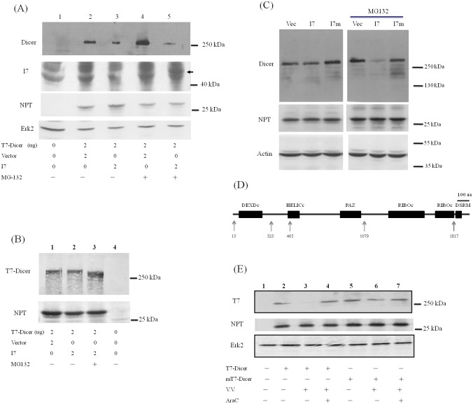
Fig 2. Cleavage of Dicer protein by I7 protease facilitates Dicer degradation.
(A) HeLa cells were mock-transfected (lane 1), co-transfected with the plasmids expressing T7-Dicer and empty vector (lanes 2 and 4) or the plasmids expressing T7-Dicer and I7 protease with V5 tag (lanes 3 and 5). Thirty-two hrs after transfection, 10 uM MG132 was also added (lanes 4 and 5). Sixteen hrs later, cell lysates were analyzed by SDS-PAGE and Western blotting with antibodies against the extreme C-terminus of Dicer protein (upper panel), V5-tag to detect the expression of I7 protease, NPT protein as the transfection control, or Erk2 protein as the loading control (bottom panel). The arrow marks the position of I7 in lane 5. (B) HeLa cells were mock-transfected (lane 4), co-transfected with the plasmids expressing T7-Dicer and empty vector (lane 1) or the plasmids expressing T7-Dicer and I7 protease (lanes 2 and 3). Thirty-two hrs after transfection, 10 uM MG132 was also added (lane 3). Sixteen hrs later, cell lysates were analyzed by SDS-PAGE and Western blotting with antibodies against the T7 tag at the N-terminus of Dicer protein (upper panel) or NPT protein as the transfection control. (C) HeLa cells were transfected with empty vector (Vec), plasmids expressing I7 protease (I7) or I7 protease containing C328A mutation (I7m). Twenty-four hrs after transfection, DMSO (left panels) or 20 uM MG132 (right panels) was also added. Twenty-four hrs after treatment, cell lysates were analyzed by SDS-PAGE and Western blotting with antibodies against Dicer (upper panel), NPT protein as transfection control (middle panel) or β-actin for the loading control (bottom panel). (D) Different functional domains in the Dicer protein. Five potential viral I7 protease cleavage sites (a.a. 13, 323, 465, 1079 and 1817) in Dicer protein are marked by arrows. (E) HeLa cells were mock-transfected (lane 1), transfected with the plasmid expressing T7-Dicer (lanes 2–4) or transfected with the plasmid expressing T7-Dicer with mutations in a.a. 1816 and 1817 (mT7-Dicer, lanes 5–7). Twenty-four hrs after transfection, these cells were either mock infected (lanes 2 and 5), or infected with VV (M.O.I. = 5) in the presence (lanes 4 and 7) or absence (lanes 3 and 6) of 100 ug araC. Twenty-four hrs later, cell lysates were analyzed by SDS-PAGE and Western blotting with antibodies against the N-terminal T7 tag of Dicer protein (upper panel), NPT protein as the transfection control or Erk2 protein as the loading control.

