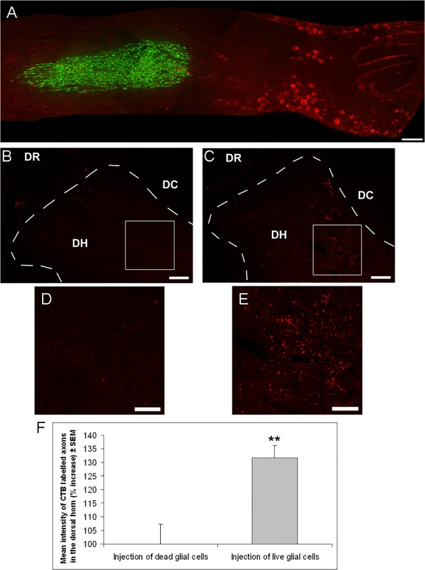
Fig 2. Transplantation of retinal glia significantly enhances DRG axon regeneration past the dorsal root entry zone.
(A) Composite photomicrograph showing GFP+ retinal glia (green) that had been transplanted two weeks earlier 1–2 mm distally to CTB labelled L5 DRG neurons (red) into the spinal nerve side in an adult rat. (B,C) Low magnification images, of the dorsal root (DR), dorsal horn (DH) and dorsal column (DC) and (D,E) high magnification images of the dorsal horn, showing CTB labelled DRG axons, after transplantation of (B,D) dead retinal glia or (C,E) live retinal glia. (F) Quantification of the percentage increase in intensity of CTB labelled axons in the dorsal horn after transplantation of dead or live retinal glia in adult rats (n = 7 rats, dead glia; n = 8 rats, live glia). Animals in A-F were sacrificed two weeks after surgery. Significant differences are indicated by asterisk (P < 0.01 = **). Scale bars: (A) 200μm; (B,C) 100μm; (D,E) 50μm

