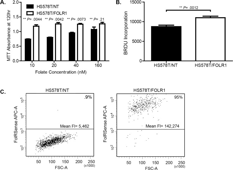
Fig 7. Folate receptor overexpression increases cell growth and folate uptake.
(A). Growth of HS578T breast cancer cells engineered to stably overexpress FOLR1 or empty vector (NT). Cells were cultured in low (0–40 nM) and super-physiological (160 nM) concentrations of folic acid and growth determined after 120 hr using the MTT assay. Error bars represent ± SD. **Statistical significance calculated by two sided unpaired t-test, assuming unequal variances. (B). BrdU incorporation in HS578T/NT and HS578T/FOLR1 cells grown in 10 nM folic acid. **Statistical significance calculated by two sided unpaired t-test, assuming unequal variances. (C). Folate uptake in HS578T/NT versus HS578T/FOLR1 cells. Briefly, flow cytometry was used to measure internalized fluorescent folate (fluorescent tagged folate agent, FolateRSense 690) after 1 hr of exposure. Dot plots show forward scatter area (FSC-A) vs. FolateRSense fluorescence (APC). The percent of cells with internalized fluorescent folate and the mean fluorescence per cell type are shown.

