Fig. 2.

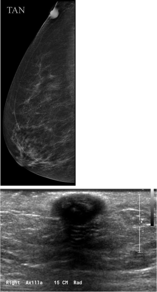
Sebaceous cyst or epidermal inclusion cyst. a Tangential view reveals that this lesion is located within in the skin. b Ultrasound confirms a hypoechoic mass within the skin

Sebaceous cyst or epidermal inclusion cyst. a Tangential view reveals that this lesion is located within in the skin. b Ultrasound confirms a hypoechoic mass within the skin