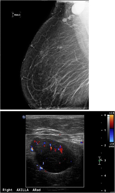
Fig. 9.

A 55-year-old African American female for screening. a MLO views of the breasts demonstrate a dense enlarged right axillary lymph node and several abnormal-appearing intramammary lymph nodes. Right axillary ultrasound confirms a large fatty replaced lymph node with non-hilar internal vascular flow. This is highly suspicious for malignancy in a patient with breast cancer; however this patient had a negative mammogram and so an ultrasound-guided core biopsy was performed for diagnosis. Final diagnosis: sarcoidosis
