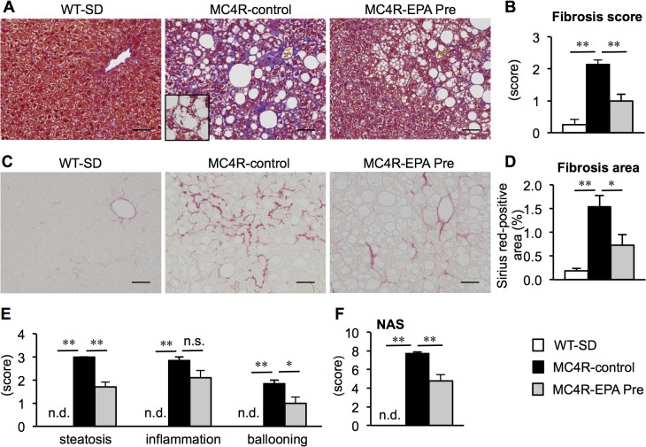
Fig 2. Effect of EPA on liver injury and fibrosis in MC4R-KO mice.
Fibrillar collagen deposition evaluated by Masson-trichrome staining (A) and fibrosis scores (B) at 24 weeks. Inset: Representative image of hepatocyte ballooning. Sirius red staining (C) and quantification of Sirius red-positive area (D). Scores of steatosis, lobular inflammation, ballooning degeneration (E) and non-alcoholic fatty liver disease activity score (NAS) (F). Scale bars, 50 μm. * P < 0.05; ** P < 0.01; n.s., not significant; n.d., not detected. WT-SD, n = 8; MC4R-control, n = 7; MC4R-EPA Pre, n = 10.

