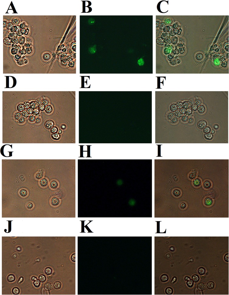
Fig 2. Immunofluorescent analysis of P247 and P523 expression in megalocytivirus-infected fish.
Peripheral blood leukocytes were collected from turbot infected with (A, B, G, and H) or without (D, E, J, and K) megalocytivirus. The cells were treated with rat antibodies against recombinant P247 (A, B, D, and E) or P523 (G, H, J, and K) and then with FITC-labeled goat anti-rat antibodies. The cells were observed under a microscope with (B, E, H, and K) or without (A, D, G, and J) fluorescence. Panels C, F, I, and L are merges of A and B, D and E, G and H, and J and K respectively. Magnification, 10×40.

