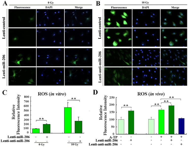
Fig 5. miR-206 regulated ROS levels in vitro and in vivo.
(A) Determination of ROS baseline levels of primary canine myocardial cells infected or not with lenti-miR-206. (B) To induce ROS, cells infected or not with lenti-anti-miR-206 exposed to 10 Gy of X-ray irradiation. Forty-eight hours after infection, the ROS levels were determined using the ROS-sensitive dye 2’,7’-dichlorofluorescein diacetate (DCF-DA). Fluorescent signals, reflecting the concentration of ROS, were measured by a fluorescence microscope under the same conditions. (C) Relative ROS levels in indicated groups of cells, as calculated by Image J analysis software (MD, USA). The normalized fluorescent signals of the cells infected with the control lentivirus were set as 100. (D) ROS levels were determined using DCF-DA in tissues. Dogs were injected with control lentivirus or the miR-206-overexpressing lentivirus. Two weeks after infection, the ROS levels in fresh tissues were measured. The level of DCF fluorescence was measured at 488 nm using a 96-well plate reader. **P < 0.01.

