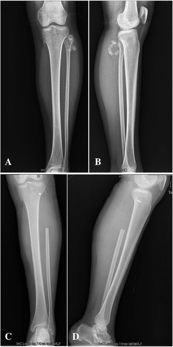
Figure 1.

Radiographs of the patient’s left shank. A, B Anteroposterior and lateral views show a bone tumor with a ‘popcorn’ and ‘smoke ring’ pattern of calcification on the posterolateral aspect of the left proximal fibula, respectively. C, D Postoperative radiographs show the absence of the left proximal fibula and an anchor inserted the lateral tibial metaphysis.
