Figure 4.

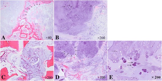
Histological appearance of tumor (H&E). A Microscopic appearance of the tissue specimen dissected from the fibula lesion shows a vaguely lobulated neoplastic hyaline cartilage separated by fibrous bands and focal myxoid change (×40). B High-power photomicrograph shows the enlarged tumor cells with moderate grade of atypia and exhibits multiple, enlarged grotesque nuclei. Mitoses can be seen (×200). C The tumor cells were seen in the Volkman canal (×200). D, E High-power photomicrograph of the tissue specimen from the needle biopsy of ilium shows a cartilaginous lesion with hypercellularity and cytologic atypia (×100, ×200, respectively).
