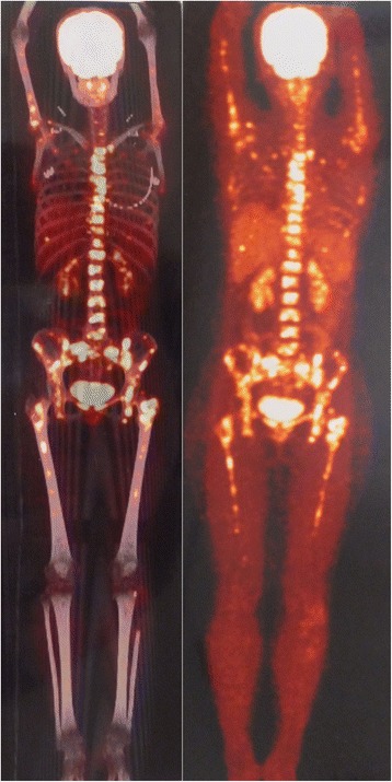
Figure 5.

PET/CT of the patient. F-18 FDG PET/CT shows multiple bone metastases especially and symmetrically in the axial skeleton and proximal extremities including bilateral femurs and humeri, with a maximum SUV of 10.8, but with no signs of local recurrence and no focal F-18 FDG uptake in the brain, head, neck, chest, and abdomen except a small nodule in the liver which was suspected as a calcification.
