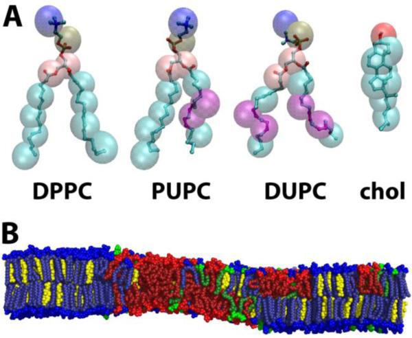
Figure 1.
Molecules used in this study. (A) CG (translucent spheres) and corresponding UA (ball-and-stick) representation of the four molecules used in this study. Purple denotes the location of double bonds. (B) Snapshot of equilibrated UA bilayer for ρ ~ 0.8, with DPPC (blue), PUPC (green), DUPC (red) and chol (yellow). Distinct regions of composition and order are observed. Molecule representations visualized in VMD version 1.9, snapshot visualized in PyMOL version 1.3.

