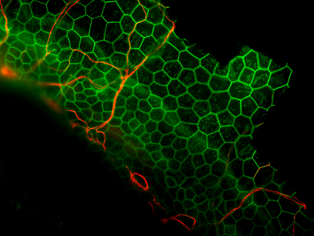
Figure 7.
Example of neurite growth toward the basilar membrane area in a deaf, neurotrophin treated ear. A whole-mount of the basal turn of the guinea pig cochlea stained for neurofilaments (red) and actin (green) and viewed with epi-fluorescence is shown. The ear was deafened with neomycin, injected with AAV. NTF-3 a week later and obtained for histology 3 months after that. The auditory epithelium does not contain differentiated hair cells or supporting cells. Instead, it is composed of flat or cuboidal simple epithelium. Nerve fibers are seen entering the epithelium and traversing the epithelial cells. This experiment was similar to that reported by Shibata and colleagues (2010) except that AAV was used in this case instead of Ad as the vector for gene therapy.

