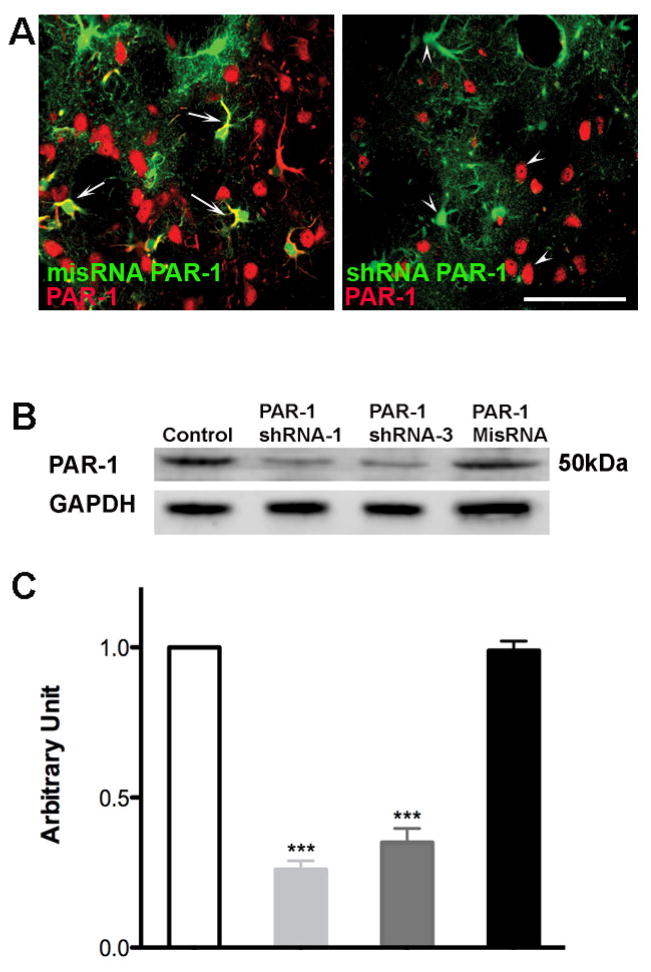
Figure 1. Effective knockdown of PAR-1 with PAR-1 shRNA lentivirus in brain sections and transfected neuronal cells.
(A) Lentivirus was injected into rat striatum. After one week the animals were sacrificed and brain sections were stained to determine the effective knockdown of PAR-1 mediated by lentivirus. Sections were stained with rabbit polyclonal antibody against PAR-1 (Alexa 594 goat anti-rabbit secondary antibody.) Pictomicrograph shows cells infected with lentivirus expressing a mis-sense RNA (misRNA) PAR-1 colocalize with PAR-1 protein indicated with arrows, demonstrating no knock down. Cells infected with shRNA PAR-1 lentivirus show no colocalization with PAR-1 receptor antibody, indicated with arrow heads. (B) Cell lysate taken from infected cells was used to determine the protein levels of PAR-1 with western blot analysis. Decreased expression of PAR-1 was observed in cells infected with PAR-1 shRNA when compared to control cells and PAR-1 misRNA infected cells. (C) Densitometry analysis shows a significant reduction in PAR-1 expression upon infection with PAR-1 shRNA lentivirus *** p<0.001.

