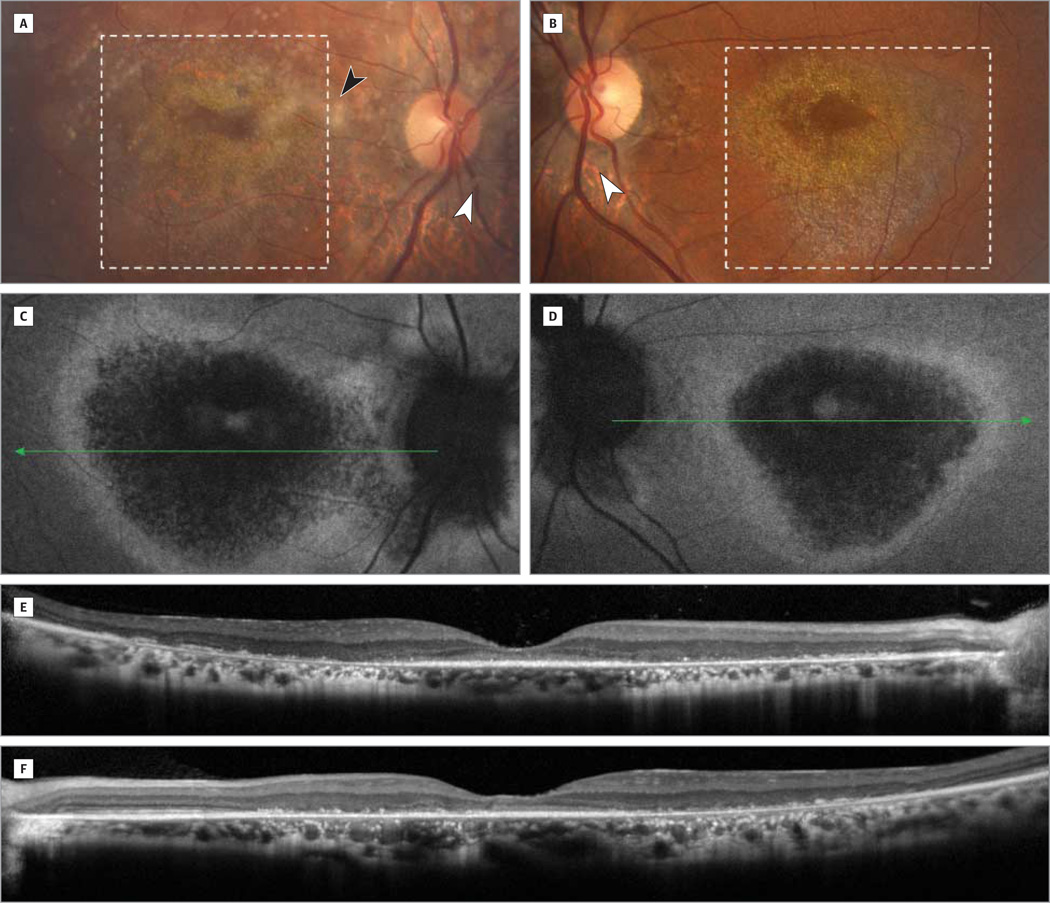
Figure 1. Color Photographs, Fundus Autofluorescence Images, and Optical Coherence Tomographic Images.
Color photographs of the right (A) and left (B) eyes demonstrate bilateral macular atrophy (dotted inset) with pale optic nerves with peripapillary atrophy (white arrowheads) and diffuse retinal pigment epithelial changes. There was also a significant amount of asteroid hyalosis (black arrowhead) in the right eye (A). These magnified images of the macula highlight the graduated atrophy corresponding to the bright deposits around the fovea in both eyes (A and B) exhibiting a hyperreflective crystalline appearance. Fundus autofluorescence images of the right (C) and left (D) eyes demonstrate hypoautofluorescent areas suggestive of central atrophy with a surrounding ring of hyperautofluorescence corresponding to lipofuscin accumulation in the retinal pigment epithelium likely secondary to incomplete degradation of photoreceptor outer segments. These autofluorescence findings correlate with retinal optical coherence tomographic imaging of the right (E) and left (F) eyes showing diffuse outer retinal atrophy bilaterally.

