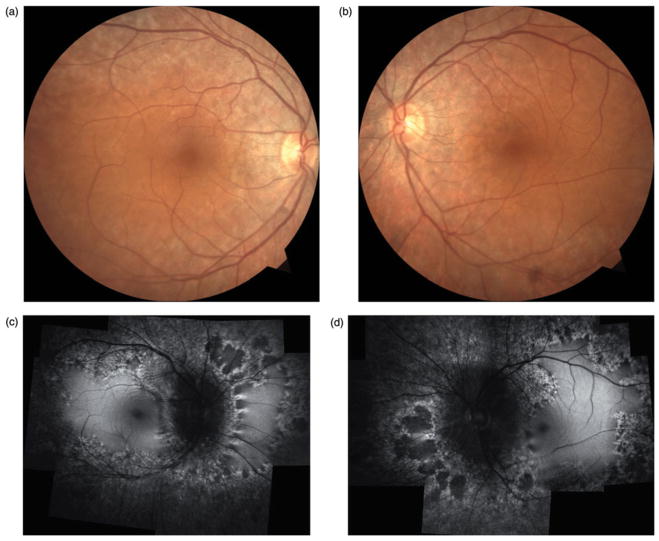
FIGURE 1.
Case 1. Fundus photographs (a & b) reveal retinal pigment epithelium atrophy along the arcades and peripapillary regions in both eyes without suggestions of intraretinal pigment migration. Fundus autofluorescence (c & d) shows extensive hypofluorescent regions along the arcades and mid-periphery. Regions of scalloped atrophy are noted nasally in both eyes.

