
FIGURE 1.
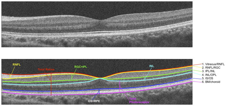
Optical coherence tomography segmentation layers in a normal eye. BM = Bruch membrane; INL = inner nuclear layer; IPL = inner plexiform layer; IS/OS = photoreceptor inner segment/outer segment junction; OPL = outer plexiform layer; RGC = retinal ganglion cell layer; RNFL = retinal nerve fiber layer; RPE = retinal pigment epithelium.