Figure 2.

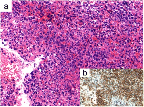
Histopathology finding of the spleen. (a) Hematoxylin-eosin stain. Aggregated large atypical cells were seen. The individual cells had chromatin-rich nuclei and relatively abundant intracytoplasmic eosinophilic inclusion bodies. (b) CD20 stain. Atypical cells were positive for CD20.
