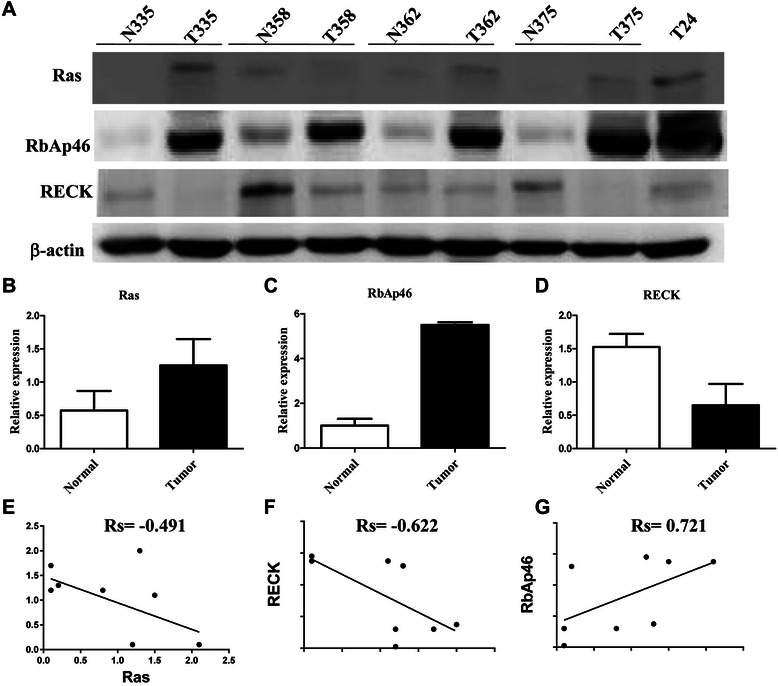
Figure 5.

Interrelationships among Ras, RbAp46 and RECK in clinical bladder cancer tissues. (A) The proteins extracted from the tissue of four clinical bladder cancer patients were evaluated by Western blotting to detect expression levels of Ras, RECK and RbAp46 using specific antibodies as indicated. β-actin was used as the internal control. N: normal tissue of bladder, and T: bladder tumor in patients. T24 cells were used as a positive control for the expression of mutant Ras and RECK. The intensity of bands for Ras (B), RECK (C) and RbAp46 (D) was quantified by a densitometer. Quantified intensity of bands was plotting using a Spearman correlation (E-G).
