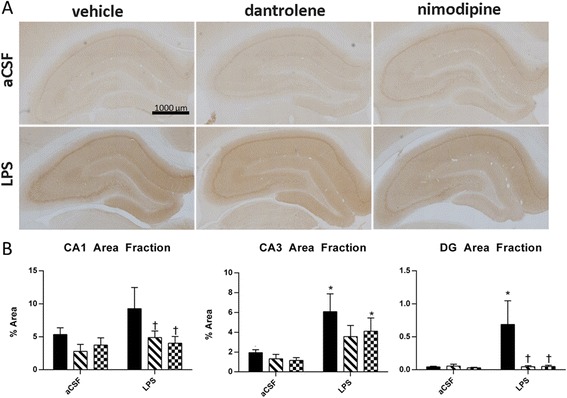
Figure 2.

Hippocampal immunohistochemistry against Arc was quantified across in brains perfused 30 min after rats were exposed to a novel context. (A) Representative slices of hippocampal Arc immunohistochemistry after 4 weeks of infusion with aCSF (top row) or LPS (bottom row) and treatment with vehicle (first column), dantrolene (middle column), or nimodipine (third column). (B) Quantification of Arc immunostaining in CA1, CA1, and DG. Data expressed as mean ± SEM. *Indicates a significant difference from treatment-matched aCSF controls, †indicates a significant difference from LPS + vehicle rats within the LPS group. Significance determined by P < 0.05. LPS lipopolysaccharide, aCSF artificial cerebrospinal fluid, DG dentate gyrus.
