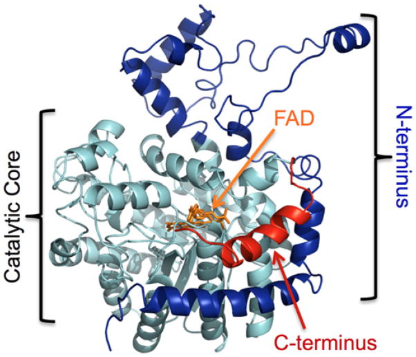
Figure 3. Homology model of PRODH2 produced by I-TASSER.
The catalytic core is highlighted in light blue. The section of the N-terminus removed in the PRODH2 157–515 construct is coloured dark blue. The C-terminal truncation is coloured red. FAD was placed into the active site based upon the structure of the PRODH domain of E. coli PutA (PDB 3E2Q).

