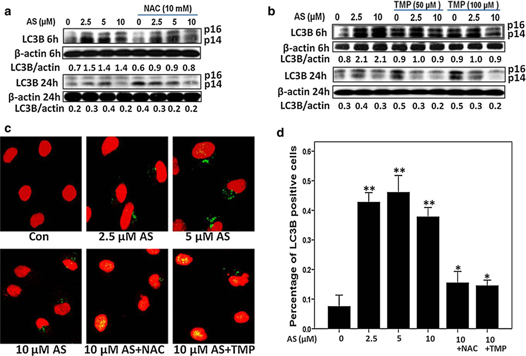
Fig. 3.
Arsenic-induced autophagy in HK-2 cells. a, b Arsenic treatment increased LC3B protein expression at 6 h, which dropped at 24 h. Both TMP (100 µM) and NAC (10 mM) inhibited arsenic-induced LC3B protein expression. β-Actin was used as loading control. c, d Induction of autophagy after 6-h arsenic exposure was visualized by LC3B staining (green), which was suppressed by 100 µM TMP or 10 mM NAC pretreatment. Values are mean ± SD (n = 3), *p < 0.05 versus Con, **p < 0.01 versus Con (color figure online)

