
BOX FIGURE 2.
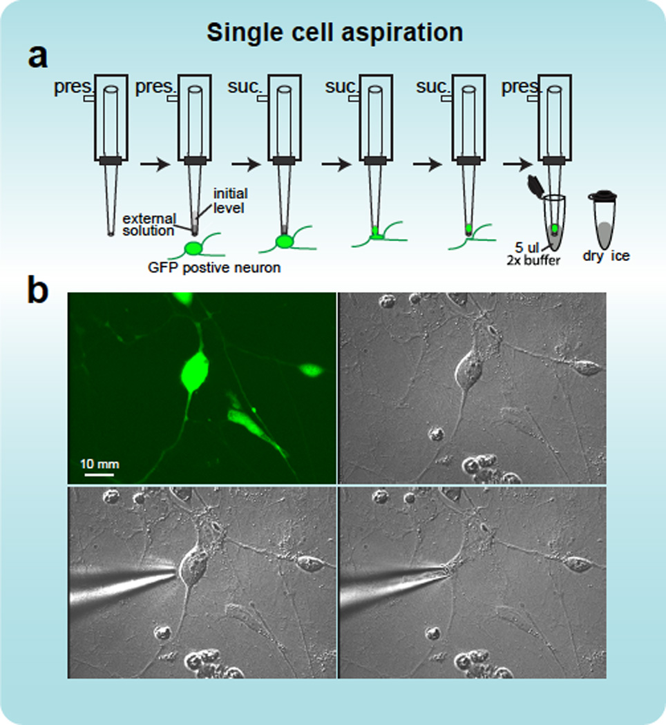
Aspiration of single cells. a, A schematic illustration of the method for collection single cells using a patch pipette. Initially, a patch pipette with a tip opening of 2 to 3 µm (~0.5 Mohm resistance) is fixed to the pipette holder, which is attached through plastic tubing to a 3 ml syringe. Slight positive pressure (pres.) is applied through the syringe while inserting the pipette into the media on the dish containing the target neuronal cells. Target cells are identified by their GFP expression and neuronal morphology, as assessed by differential-interference contrast (DIC) imaging. The positive pressure driven through the pipette while it is lowered into the solution reduces the influx of liquid into the pipette due to capillary action. Under visual direction through the microscope, a micromanipulator is used to position the pipette close to the cell body. Once the tip touches the cell, the cell will normally adhere to the tip, at this point the pressure is relieved, and slight suction (suc.) applied, enabling gradual aspiration of the cell into the pipette tip. The pipette is then retracted, and while positioning it in a pre-prepared PCR tube with 5 µl reaction buffer, the syringe is used to apply positive pressure to expel the cell into the solution in the tube. b, Fluorescent and DIC image of the collection of an iN cell as described.