Figure 1.
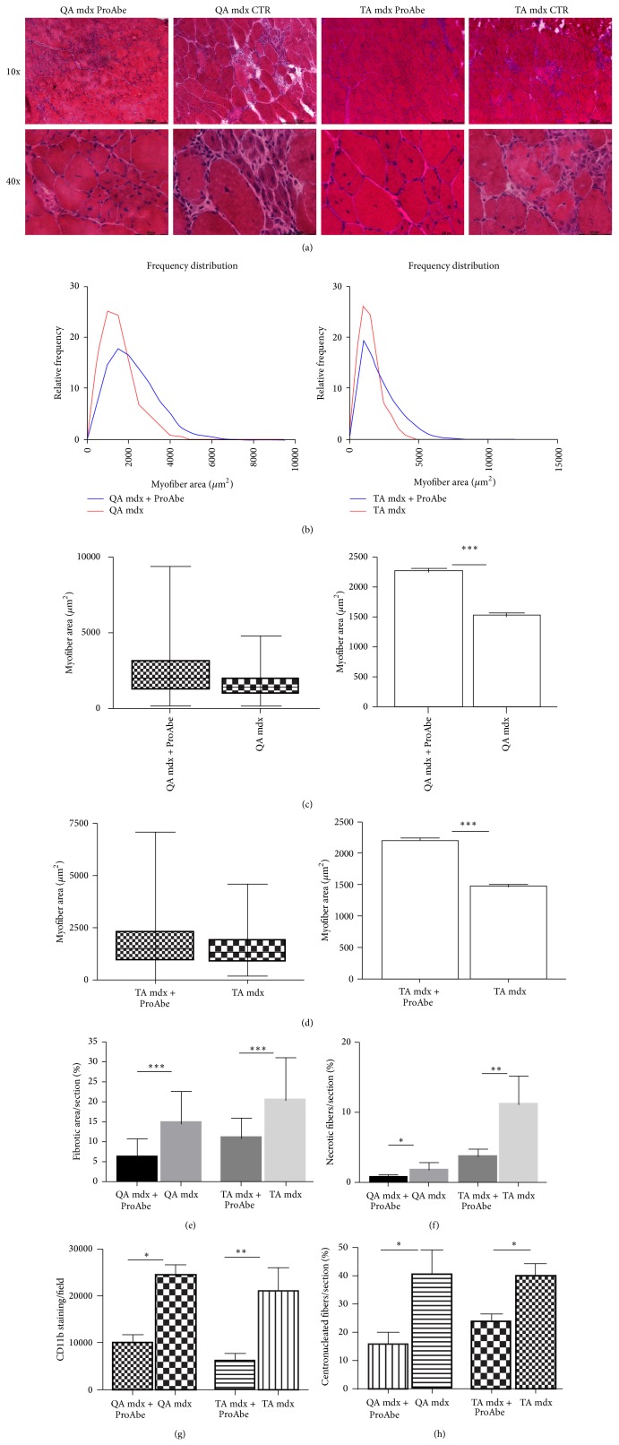
H&E analysis of treated and untreated mice. H&E staining was performed on 10 μm-thick frozen sections from TA and QA muscles (a). In treated mice reduced signs of muscular wasting were observed in comparison to untreated mice. In particular reduced inflammatory infiltrates between myofibers, reduced fat deposition, and reduced necrotic fibers were assessed. In both treated and untreated mice small centronucleated regenerating fibers were observed. For each muscle analyzed with H&E staining we showed the myofiber area and their distribution frequency (b). The curve of TA/QA muscles of treated mice shifted to the right related to untreated mice, thus demonstrating an increase in myofiber area of treated mice (CSA QA: 25° percentile of treated mice 1221,26 and of untreated mice 903,152; CSA TA: 25° percentile of treated mice 1120,39 and of untreated mice 874,068; CSA QA: 75° percentile of treated mice 3107,04 and of untreated mice 1946,06; and CSA TA: 75° percentile of treated mice 2978,59 and of untreated mice 1883,39). Moreover, we indicated the coefficient of variance (graphs showing Min and Max values and mean + SEM) ((c) for QA and (d) for TA). Amelioration of dystrophic phenotype following ProAbe treatment was demonstrated by decrease of fibrosis by measuring area of connective tissue (AM) (e), of the percentage of necrotic fibers (f), of macrophage infiltration area (CD11b staining) (g), and of the number of centronucleated myofibers per section (h).

