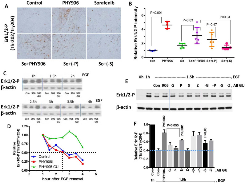
Figure 6. Effect of PHY906, Sorafenib (So), Sorafenib+PHY906 (So+PHY906), Sorafenib+PHY906 deleted P (So+(-P)), and Sorafenib+PHY906 deleted S (So+(-S)) on Erk1/2 phosphorylation of HepG2 tumors in NCR nude mice.
(A) Immunohistochemistry staining for phosphorylated Erk1/2 (Thr202/Tyr204) in HepG2 tumor sections after the drug treatment for 96 h. (B) Quantification of immunohistochemistry staining of Erk1/2 phosphorylation using imaging software. (Each spot represents a mean of the intensity of brown color from 5 views of a tumor section; number of animals is 5). (C) Western blotting analysis for the effect of PHY906 or E.coli β-glucuronidase treated PHY906 (500 μg/ml) on dephosphorylation rate of Erk1/2 in HepG2 cells following stimulation with EGF (50 ng/ml). β-actin was used as the loading control for normalization. Cropped blots are used in this figure and they have been run under the same experimental conditions (please see the full-length bolts in Fig S16A) (D) Quantification of the Western blot results for the phosphorylated Erk1/2 (Thr202/Tyr204). (E) Western blotting analysis for the effect of E.coli β-glucuronidase treated PHY906 (500 μg/ml), equivalent concentration of single herbs (G, P, S, Z), or equivalent concentration of a one herb deleted formula (-G, -P, -S, -Z) on dephosphorylation rate of Erk1/2 in HepG2 cells following stimulation with EGF (50 ng/ml). Cropped blots are used in this figure and they have been run under the same experimental conditions (please see the full-lenght bolts in Fig S16B) (F) Quantification of the Western blot results for the phosphorylated Erk1/2 (Thr202/Tyr204). Details of experimental procedures are given in Materials and Methods.

