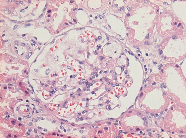
Fig. 1.
Detail of a glomerulus showing a segmental area of scarring located at the tip with adhesions to the Bowman's capsule. Foam cells are present within the sclerosed capillary loops. The remaining glomerular tuft shows open capillary loops without endocapillary hypercellularity (HE staining and ×200).

